> Vet Referrals > Services Offered > Orthopaedic Surgery

Orthopaedic Surgery

Including Cruciate Ligament Disease in Dogs


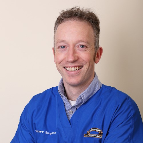
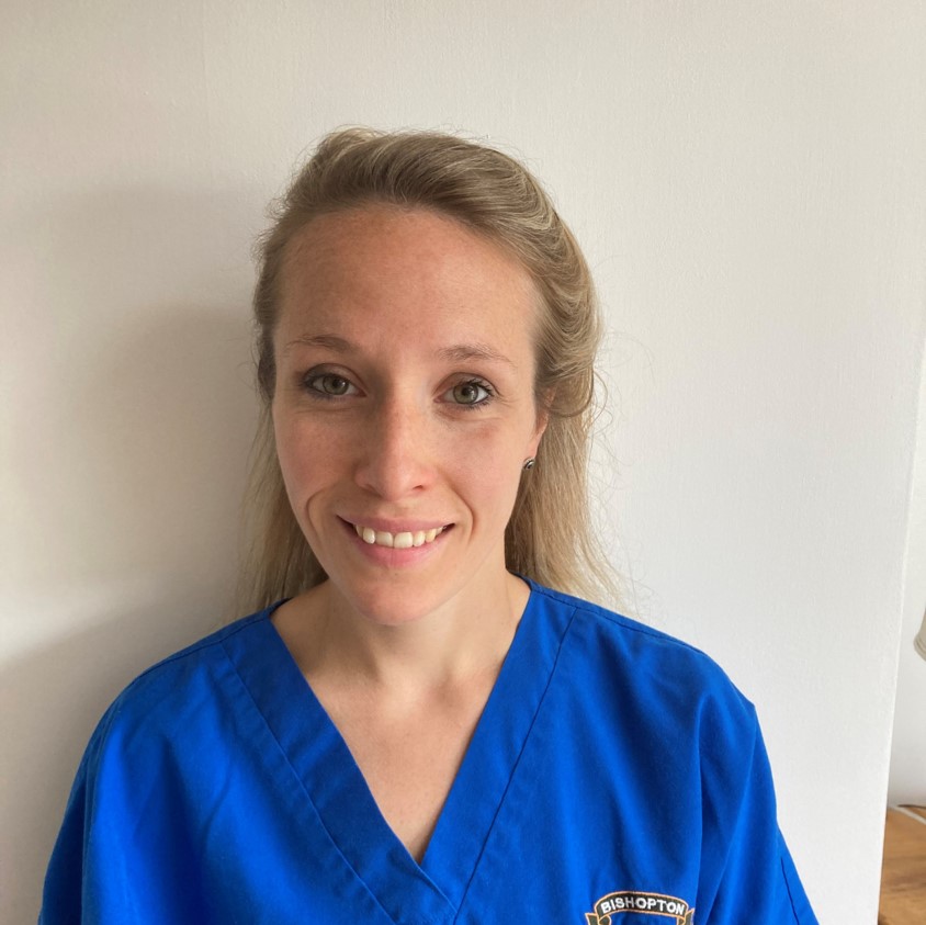
Our surgical team is led by Ian McClive MA VetMB CertSAS MRCVS, RCVS Advanced Practitioner in Small Animal Surgery. Ian is supported by vet Laura Pearce BVetMedSci (Hons) BVM BVS PgC(SAS) MRCVS RCVS, Advanced Practitioner in Small Animal Surgery

Our surgical nursing team is supported by Claire Dennis and Laura Hagston. 

We perform a wide range of orthopaedic procedures including but not limited to the following:

  • Cruciate ligament injury (repair by TTA)
  • Fracture repair
  • Arthrodesis
  • Medial Patellar Luxation correction
  • Lateral humeral condylar fracture
  • Coxo-Femoral Luxation (dislocation of the hip joint)

We believe in sensible fixed pricing for many of these more complicated procedures enabling owners to budget for their pet's care. Our prices include initial consultation, radiographs, blood work, surgical procedure, associated medications, hospitalisation, two post-operative checks, initial follow-up radiographs at 4 – 6 weeks if indicated, any post-surgical complications within the first 6-months and VAT.

Cruciate Ligament Disease in Dogs

Background

There are two cruciate ligaments in the knee (or ‘stifle’ as we call it in dogs). Cruciate disease (affecting the cranial cruciate ligament) is one of the most common orthopaedic conditions seen in dogs. In people cruciate ligament ruptures are typically seen as acute sporting injuries in footballers and skiers for example. In dogs, it is a little different and presents as a more chronic degenerative condition. The ligament degenerates and gets weaker with time, and at some point, will start to tear.

Diagnosis

The diagnosis is often made on palpation/manipulation of the stifle, although in some dogs this may require sedation. X-rays will show signs of osteoarthritis (OA or ‘arthritis’). Early cases may be less easy to diagnose, sometimes requiring exploratory surgery/arthroscopy to visually examine the cruciate ligament.

Treatment

Some small dogs (less than 15kg) may do well with a period of rest and anti-inflammatories alone, although surgery is generally considered to offer a quicker and more reliable recovery. Larger dogs are less likely to do well without surgery and so surgery is always advised. Numerous stabilisation techniques have been described. The most successful techniques involve placing a restraining suture around the outside of the joint to try to replicate the function of the torn ligament (known as extracapsular stabilisation) or changing the geometry of the tibia to counteract the forces that are responsible for the instability.

We are advising to treat your pet’s cranial cruciate ligament failure surgically using a geometry changing technique known as the Modified Maquet Procedure (MMP). The operation is based on a technique developed almost 50 years ago for use in human knees by Dr Maquet, a Belgian orthopaedic surgeon. The operation works by redirecting the force generated by the large quadriceps muscles to compensate for the failed cruciate ligament. This is achieved by cutting free, and moving forward, the part of the tibia (the tibial tuberosity) attached to the quadriceps muscle. The bone cut is called an osteotomy and the osteotomy is stabilised using a modern orthopaedic implant material called Orthofoam. The porous titanium Orthofoam promotes remarkably rapid bone ingrowth and healing and this is key to the reduced convalescence and minimal pain seen with MMP surgery.

Successful recovery after knee surgery, no matter which procedure has been used, requires a period of controlled activity. Compared to other procedures, MMP causes less discomfort and while a comfortable, pain free patient is obviously a good thing, many dogs are tempted to use the operated leg too much, too soon. No matter how comfortable and confident your pet is feeling in the days after their MMP operation, it is absolutely essential that running, jumping, and general “rough and tumble” with other pets is avoided for the first 6 weeks or so. The bone must be given time to heal adequately and too much strain placed on the osteotomy too early can result in stress fracture or implant failure and while this is rarely catastrophic, the ensuing complication may be painful and will certainly delay the recovery. The operation works by redirecting the force generated by the large quadriceps muscles to compensate for the failed cruciate ligament. This is achieved by cutting free, and moving forward, the part of the tibia (the tibial tuberosity) attached to the quadriceps muscle. The bone cut is called an osteotomy and the osteotomy is stabilised using a modern orthopaedic implant material called Orthofoam. The porous titanium Orthofoam promotes remarkably rapid bone ingrowth and healing and this is key to the reduced convalescence and minimal pain seen with MMP surgery.